Identifying Your Pain Source

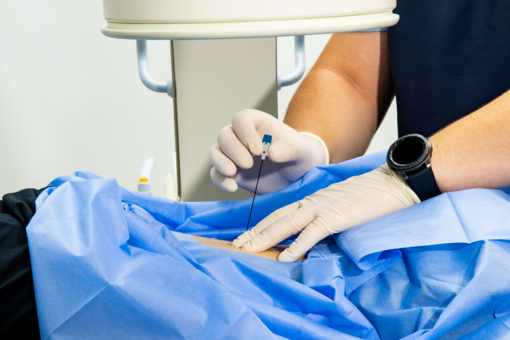
Imaging-guided diagnostic blocks use targeted anesthetic injections to pinpoint your exact source of pain. By isolating specific structures, we can distinguish between multiple potential pain generators to ensure your treatment plan is as accurate as possible.

fluoro spine procedure
barbotage shoulder

How it works.

We use real‑time ultrasound or fluoroscopy (X‑ray) guidance to place a small amount of local anesthetic directly into or around the suspected pain source. If your symptoms significantly improve or resolve within minutes to hours, we’ve confirmed that structure as the primary pain generator. If little or no relief occurs, we can rule out that source and investigate other possibilities.

What to expect.

Duration

Most diagnostic blocks take 2–10 minutes and can often be performed during your office visit.

Immediate feedback

You will know within minutes if the injection site is the source of your pain, with relief typically lasting for several hours.

Minimal discomfort

Your comfort is our priority. We use a local anesthetic to make the quick needle insertion as gentle and easy as possible.

Activity trial

We may ask you to gently reproduce movements that typically cause pain to assess the block’s effectiveness.

Documentation

We’ll record your pain levels before and after the injection to guide treatment decisions.

Safety

While this procedure is considered very safe, common side effects include temporary numbness or mild bruising. Although rare, risks such as infection, tendon or nerve injury, or a hematoma at the injection site can occur. We will discuss personalized precautions based on your medical history and the specific treatment area.

Common conditions and pain sources evaluated

Hip and groin pain.

  • Hip Osteoarthritis: Distinguish pain within the hip joint from pain referred from the lumbar spine or SI joint.
  • FAI: Differentiate mechanical impingement within the hip from pain referred from a hip flexor tendon or the lumbar spine.
  • Labral Tear: Isolate hip labral pain from extrinsic SI joint or lumbar spine referral sources.
  • Hip stabilizer Tendinopathy: Differentiate hip stabilizer tendon pathology from referral pain originating in the intra-articular hip, SI joint, or lumbar spine.

Shoulder pain.

  • Glenohumeral Arthritis: Distinguish true joint pain from cervical spine, rotator cuff, or subacromial origins.
  • AC Joint Arthritis: Isolate focal AC joint pain from referred cervical, rotator cuff, or subacromial symptoms.
  • Rotator Cuff Tendinopathy: Localize from specific rotator cuff tendons from pain originating within the shoulder or AC joint or referred from the cervical spine.
  • Biceps Tendon Pathology: Differentiate bicipital pain from referred cervical, intra-articular, AC joint, or subacromial sources.

Knee pain.

  • Intra‑articular knee pathology: Distinguish true knee joint pain from referred pain from the hip or lumbar spine.
  • Knee tendinopathy: Differentiate pain originating from a tendon (quadriceps or patellar) versus pain originating within the knee joint.
  • Pes anserine syndrome: Isolate pain originating from the pes anserine tendon vs a medial meniscus tear, the insertion of the MCL, or from patellar tendinopathy.
  • Insertional IT Band Syndrome: Localize pain from the IT band insertion vs pain associated with hamstring tendinopathy, patellar tendinopathy, or the lateral collateral ligament (LCL) insertion.

Back and neck pain.

  • Cervical Facet Pathology: Differentiate primary facetogenic pain from cervical discogenic pain, shoulder pathology, or myofascial referred pain.
  • Occipital Neuralgia: Distinguish true neuralgia from referred pain originating in the upper cervical facets or tension-type headaches.
  • Lumbar Facet Pathology: Isolate facetogenic sources from lumbar discogenic pain, sacroiliac (SI) joint referral, or intra-articular hip pathology.
  • Sacroiliac (SI) Joint Dysfunction: Localize pain to the SI joint versus referred pain from the lumbar spine, hip joint, or piriformis syndrome.

Nerve‑related pain.

  • Piriformis syndrome: Differentiate gluteal pain caused by piriformis syndrome from lumbar radiculopathy, sacroiliac (SI) joint dysfunction, or intra-articular hip pathology.
  • Carpal tunnel syndrome (CTS): Confirm median nerve compression at the wrist vs. proximal median neuropathy (e.g., Pronator Syndrome) or C6–C7 cervical radiculopathy. 
  • Cubital tunnel syndrome: Differentiate ulnar neuropathy at the elbow from C8–T1 cervical radiculopathy or distal ulnar nerve entrapment at Guyon’s canal.
  • Tarsal tunnel syndrome: Distinguish posterior tibial nerve compression at the ankle from an S1 lumbar radiculopathy.
  • Meralgia Paresthetica: Differentiate lateral femoral cutaneous nerve (LFCN) compression from an L2/L3 lumbar radiculopathy.

Diagnostic blocks provide clarity when imaging studies are inconclusive or when multiple pain generators are possible. This precision helps us recommend the most targeted and effective treatment plan for your specific condition.

The Science Behind Our Treatments.

What to know more about our evidence based approach? Explore the links below for more information.

female shoulder pain

Study 1

Comparison of ultrasound-guided versus blind glenohumeral injections: a cadaveric study.

Study 2

Comparison of ultrasound-guided (USG) versus palpation-guided AC joint injections: 2 cadaveric studies.

Shoulder ultrasound
fluoro spine procedure

Study 3

Study demonstrating accuracy, safety and clinical utility of ultrasound-guided cervical facet injections.

Get Back to What You Love

Schedule a consultation to explore non-surgical treatment options that align with your active lifestyle.